Awake-surgery
Gliomas : represent more than 50 % of all intracerebral tumors and 2/3 are malignant. (WHO grade IV ) and malignant astrocytoma (grade III) represent almost the totality of the malignant gliomas, while the rest consists of astrocytoma, benign (grade I) or low-grade (grade II). Gliomas, although benign, tend to infiltrate the surrounding brain tissue, often making the radical removal very difficult.
The oligodendroglioma and the ependymoma are other glial tumors, with relatively more favorable course.
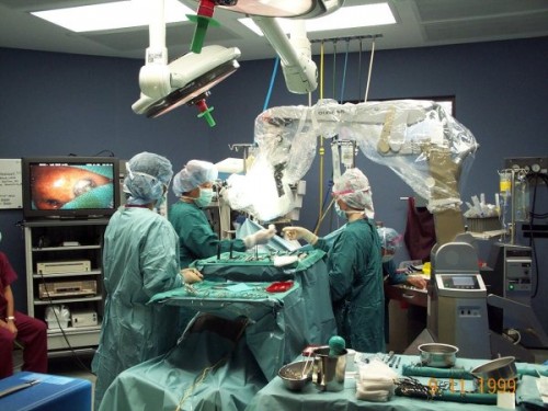
The purpose of awake surgery (AWS) is to remove as much tumor as possible in gliomas of low and high grade, without causing additional and permanent neurological damage, by using techniques of intraoperative cortical and subcortical stimulation.
The hemispherical glial tumors are frequently located near or within functional cortical and subcortical areas (sensory-motor ways, areas of language, supplementary motor area, the internal capsule, corona radiata, uncinatus fasciculus, etc). Consider also the great variability among individuals in the localization of the areas of language, that goes beyond the classical division in the anterior area, in charge of the production of spoken language (the back of the inferior frontal gyrus) and the back area, responsible for the understanding of language (the back area around the sylvian fissure of the temporo-parietal cortex)
The functional MRI (fMRI) is a technique useful to identify areas of speech, but not always completely reliable. AWS enables you to locate their distribution in temporal (middle and superior gyrus), posterior frontal and parietal lobes, and therefore to safe them.
Preoperative evaluation :
- Neurological status: presence of any motion and speech damage;
- In patients with moderate or severe dysphasia the “mapping” is impossible. In these cases, you can choose to avoid surgery or to make a
- Preoperative functional MRI.
Intra-operative precautions :
- Place the patient on the operating table on pillows, in a comfortable position, in the lateral decubitus position, in a manner he can look at the screen of a computer;
- Standard intubation or laryngeal mask airway guided by fiberoptic;
- Local anesthesia (skull clump, skin incision, opening of the meninges)
Techniques of “Mapping”.
Once the patient is awakened and extubated proceed to the cortical mapping techniques :
- Mapping of the motor cortex with monopolar or bipolar electrode connected to a stimulator with radio frequency from 2 mA to 16 mA: the stimulated lower rolandic area determines responses on the face and hands level;
- The preoperative antiepileptic and monitoring of the patient’s face to prevent focal intraoperative motor seizures: in this case, irrigation with cold Ringer solution;
- Identification of “essential” speech areas: areas whose stimulation induces mistakes (2 or 3 mistakes evoked every 3 attempts) are very reliable (p <0.005);
- Mapping of the speech cortex: you make nominate the patient objects shown on the screen of a computer, changing every 4 seconds, stimulating and controlling 2 or 3 times, marking the areas with cotton pads;
- Identification of Broca’s area: during the stimulation is observed a break in counting the numbers progressively without oro-pharyngeal movements (“speech arrest”) Usually this area is immediately in front of the motor cortex for the face;
- The identification of at least one essential area for the speech is necessary to obtain reliable data;
- Generally small focal areas are identified (1-2 cm ²), at least 1 at the posterior inferior frontal level and 1 or 2 at the level of the temporal lobe of the dominant hemisphere, often with defined margins;
- The most important factor to prevent postoperative speech deficits and to maintain the integrity of the speech as long as possible is the distance between the resection margin and the area of the mapped speech area: the one centimeter rule indicates an increased risk of permanent speech deficits for resection approaching at less than one cm from identified area;
- Also important is the preservation of the cerebral vascular system;
- Once started the removal of the tumor, also motor descending and sensory ascending pathways can be identified with the same parameters of stimulation;
- The sensory-motor pathways and the speech connection can be followed also at subcortical level, the internal capsule, brainstem and in the uncinate fasciculus;
- It should also be always reminded about the importance of the supplementary motor area (superior frontal gyrus), which when damaged can cause aphasia and/or contralateral hemiparesis generally transitory.
Anesthesia asleep-awake-asleep
Once the mapping of the speech has been completed, the patient is re-intubated by the endotracheal route with a fast track, a fiber optic laryngoscope or a laryngeal mask and the removal of the tumor continues under general anesthesia until the end of intervention, for greater patient comfort.
A postoperative control in a protected environment (Neurosurgical Intensive Care) is indicated in all cases for 24-48 hours. It is preferable to maintain high plasma levels of antiepileptic drugs and perform therapy with intravenous steroids for 3-5 days. It is also indicated a MRI monitoring with and without contrast within 72 hours after surgery to evaluate the surgical trauma and the residual tumor.
The motor and speech rehabilitation is indicated in cases with a transitional postoperative deficit.
Contraindications :
- Severe preoperative motor and speech deficits ;
- Insufficient cooperation of the patient (age, confusion, anxiety …);
- Problems with airways (opening of the mouth, retrognathia …) and pulmonary diseases (COPD, obesity);
- The insufficient myelination before 3-4 years precludes the direct cortical stimulation.
Conclusions
AWS “maximizes” the extent of the removal of astrocytomas and brain gliomas in eloquent areas, improves the quality of life and extends survival. In fact, the cortical and subcortical important areas can be localized within the tumor and function normally, the fact that entails the risk of permanent deficit of the function during the removal of the tumor, even if it remains in its interior.
The preoperative mapping with fRMN and PET is very useful but not sufficiently reliable. Therefore, the mapping with an intraoperative cortical stimulation is very useful in eloquent areas and the awake patient allows us to identify the precise locations of speech and sensorimotor areas. The anesthetic method asleep-awake-asleep minimizes the discomfort for the patient.
References :
Attachments :


